|
|
|
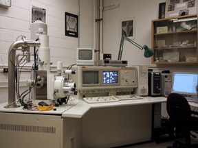
JEOL 6400 SEM |
|
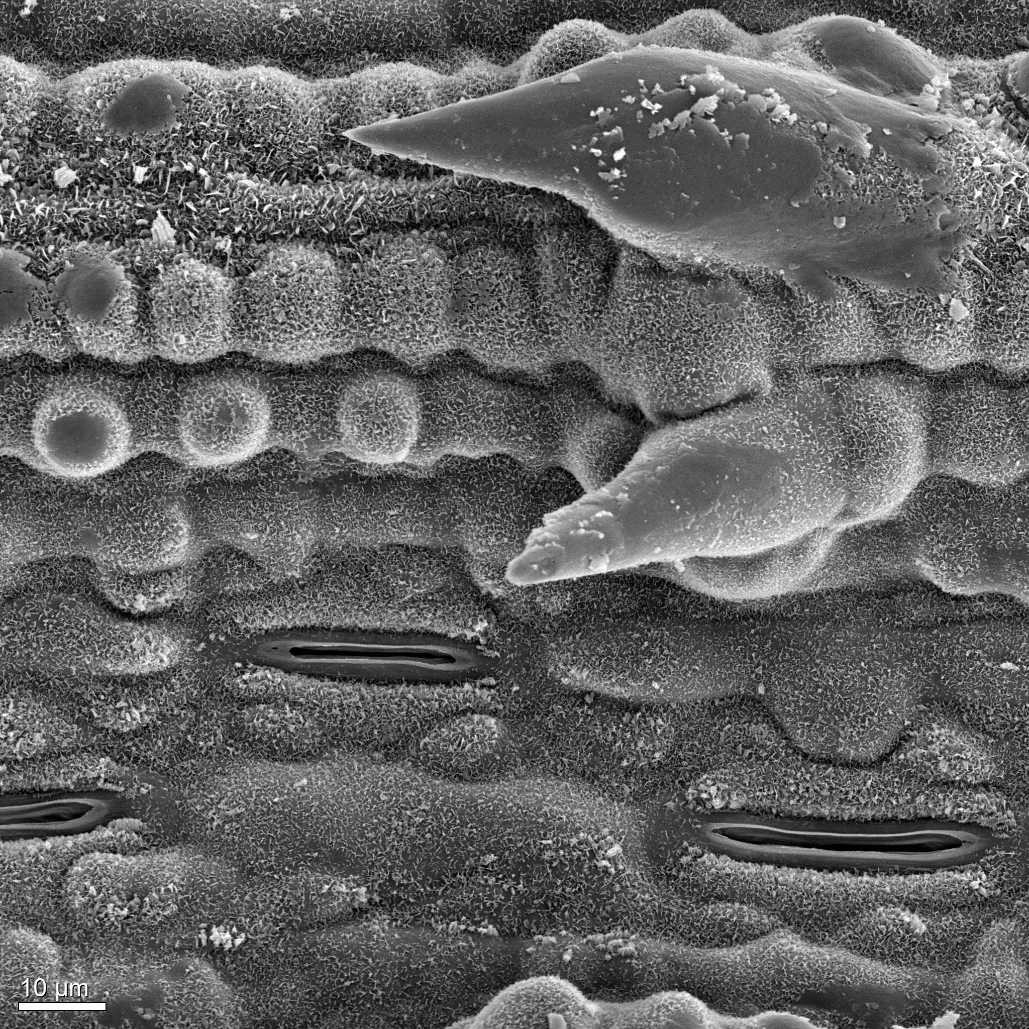
Image: Class of Biology 3331, UNB |
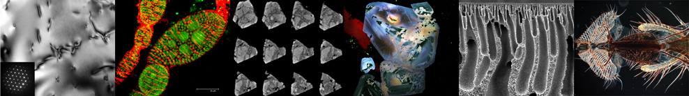
SCANNING ELECTRON MICROSCOPY
The SEM can be used to examine
surface details of solid materials. Internal surfaces
can be exposed by sectioning or fracturing. It is
equipped with an energy dispersive spectrometer
which permits qualitative and quantitative compositional
analysis. Image analysis software permits detection,
measurement and analysis of features of interest.
A cryo-stage offers the examination of frozen, hydrated
samples while a dynamic tensile stage gives an in-situ
examination of stress on materials.
IMAGING TECHNIQUES
-
Secondary electron imaging
(morphology and surface topography)
-
Backscattered electron imaging
(compositional contrast and phase distribution)
-
Digital image collection, enhancement
and analysis
- Cathodoluminescence imaging
ANALYTICAL MODES
- Elemental recognition and phase identification
- Quantitative compositional analysis
- Digital x-ray maps and linescans
- Analysis of particle samples
INSTRUMENTATION
JEOL JSM6400 Digital SEM with:
- Geller dPict digital image acquisition software
- Emitech K1250 Cryo-SEM system
- EDAX (Genesis) Energy Dispersive X-ray Analyzer
- Gatan Microtest 5000 dynamic testing stage
- Gatan ChromaCL Cathodoluminescence imaging system
|
|
|
|